Fig. 7.
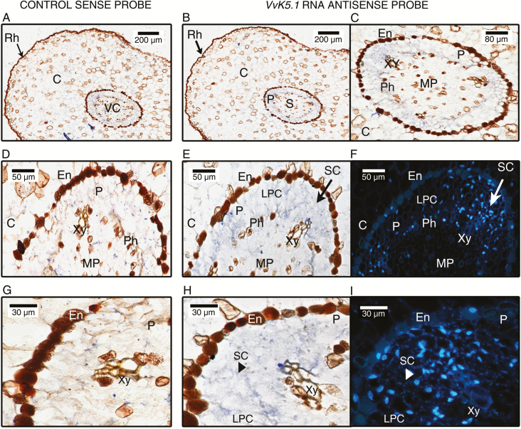
In situ localization of VvK5.1 transcripts in roots of rooted canes. Equatorial sections were hybridized with VvK5.1 RNA sense probe as negative control (left column: A, D, G) or with VvK5.1 RNA antisense probe (two right columns: B, C, E, F, H, I). Sections hybridized with VvK5.1 sense probe did not show any blue staining at the different magnifications (A, D, G). In contrast, positive signals were observed in the stele with VvK5.1 RNA antisense probe (B, C, E, H). The blue signals were located in the phloem in the small (SC) and large parenchyma cells (LPC) of the pericycle (C, E, H). A weaker signal was also detectable in the phloem. (E) and (H) were observed by fluorescence microscopy after DAPI staining to visualize nuclei and the different cell density of the pericycle (F, I). Rh, rhizodermis; C, cortex; En, endoderm; P, Pericycle; PM, medullary parenchyma; Xy, xylem; Ph, phloem; SC, small cells; LPC, large parenchyma cells.

