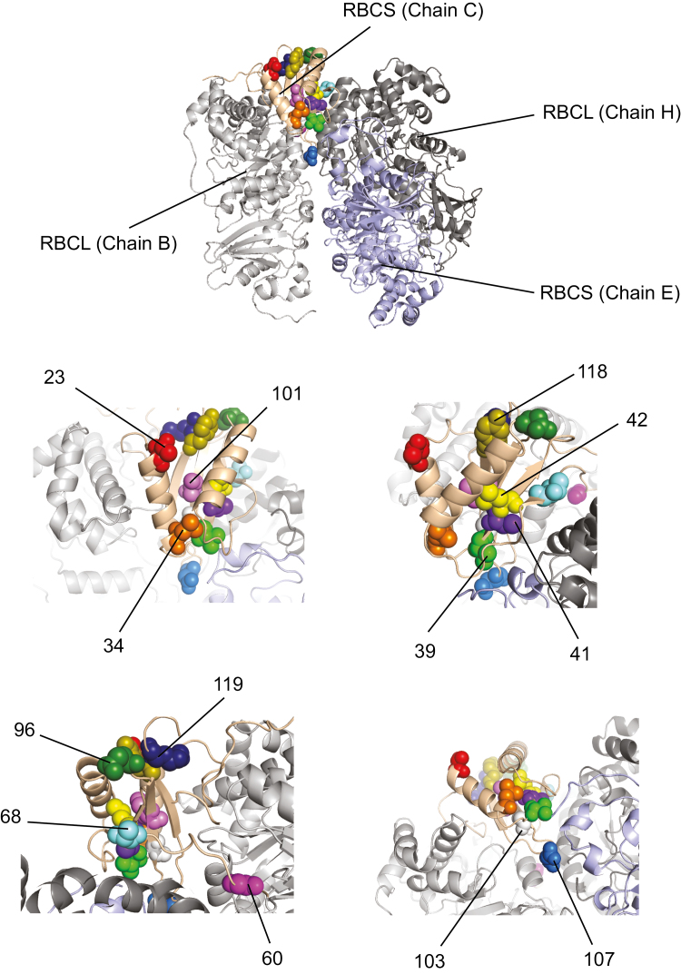
Fig. 3.
RBCS residues under positive selection. Thirteen positions of rbcS showed strong signals of positive selection (see also Table 1). We plotted corresponding amino acid residues to RuBisCO structure of Spinacia oleracea (1RCX of Protein Data Bank; Taylor and Andersson, 1997). The light pink cartoon ribbons indicate RBCS chain C of 1RCX. The light grey, light blue, and dark grey cartoon ribbons indicate RBCL chains B, E, and H of 1RCX, respectively. The positions of RBCS under positive selection are shown as spheres in different colours. The upper panel shows the overview of the positions under positive selection. The other four panels show the zoom view of each sphere.

