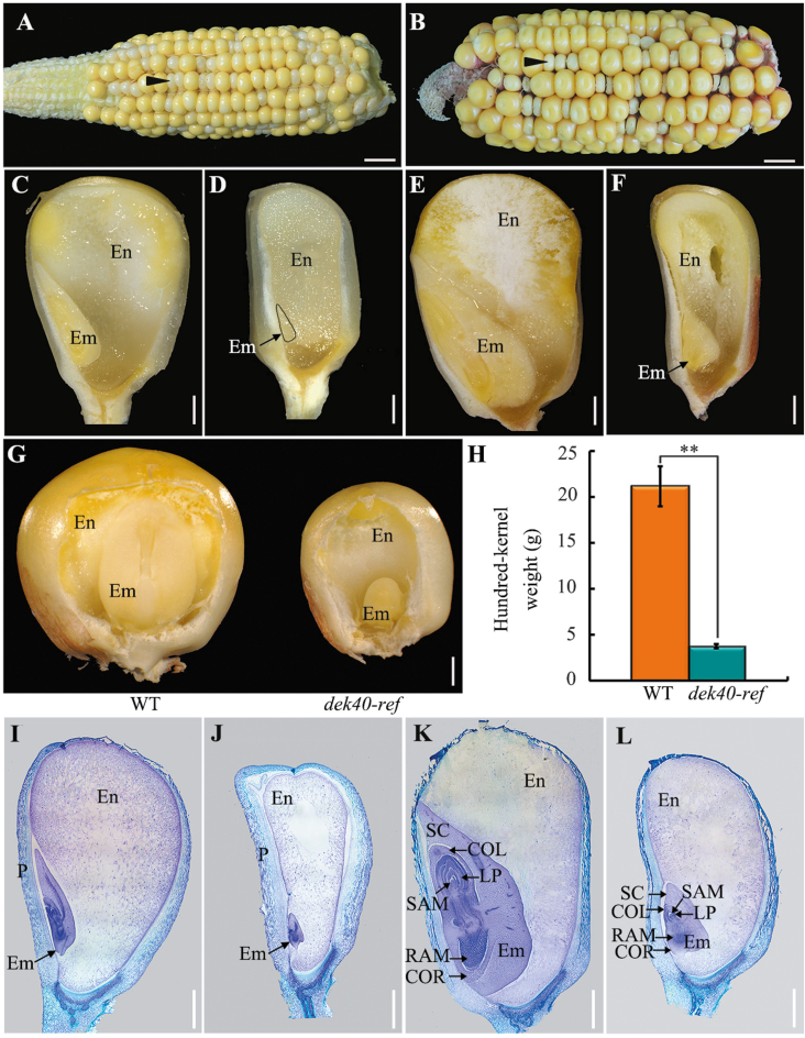
Fig. 1.
Phenotypic characterization of dek40-ref kernels. (A and B) The self-pollinated dek40-ref heterozygote ears at 15 DAP (A) and 25 DAP (B). The arrowhead indicates one of the dek40-ref kernels. Scale bars=10 mm. (C–F) Developmental comparisons of dissected WT and dek40-ref kernels at 15 and 25 DAP. (C and E) WT kernels at 15 and 25 DAP. (D and F) dek40-ref kernels at 15 and 25 DAP. Scale bars=1 mm. (G) The front view of mature WT and dek40-ref kernels whose partial pericarp was removed to display an embryo. Scale bars=1 mm. (H) Comparison of the 100-kernel weight of randomly selected mature WT and dek40-ref kernels. (I–L) Histological analysis of WT and dek40-ref kernels at 15 and 25 DAP. (I and K) WT at 15 and 25 DAP. (J and L) dek40-ref kernels at 15 and 25 DAP. Scale bars=1 mm. En, endosperm; Em, embryo; P, pericarp; LP, leaf primordia; RAM, root apical meristem; SAM, shoot apical meristem; SC, scutellum; COL, coleoptile; COR, coleorhiza.

