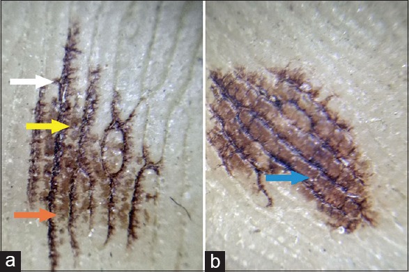
Figure 2.

Dermoscopy from light brown (a) and dark brown (b) macule: Wet contact dermoscopy of the lesion revealed dark brownish linear pigmentation along the dermatoglyphic lines (parallel furrows) with feathering (White arrow). In addition, there were blotches of light brown pigmentation interspersed between the lines (Orange arrow), which tended to coalesce giving a diffuse appearance (Blue arrow) in the darker lesions (b). Pigmentation was also accentuated around sweat pores (Yellow arrow) (Mode– non-polarized hand held dermoscope, magnification – 10×)
