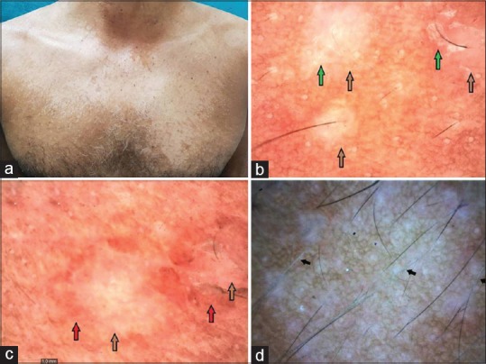
Figure 1.

Hypopigmented pityriasis versicolor: (a) Adult patient with hypopigmented variant of pityriasis versicolor involving trunk. (b) Showing primary folliculocentric lesions with reduced pigmentary network (brown arrow) with scaling at the border of the primary lesion (green arrow) (Dinolite AMZT 73915, Edge 3; magnification ×50, polarizing mode). (c) Shows similar changes with a contrast halo (hyperpigmented) around the primary lesion (red arrow) (magnification ×50). (d) Hypopigmentation of the involved hair follicle as a result of follicular invasion of the yeast marked with black arrow (magnification 50×, polarizing mode)
