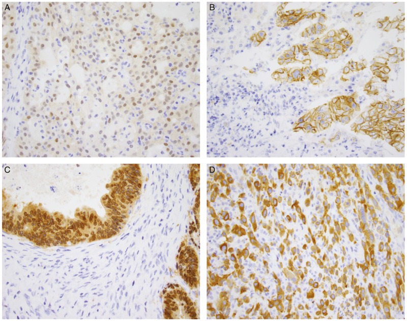
Figure 1.
Patterns of immunohistochemical staining in NTRK fusion-positive tumours. (A) Secretory carcinoma of the salivary gland with an ETV6-NTRK3 fusion shows weak to moderate nuclear and cytoplasmic staining. (B) Intrahepatic cholangiocarcinoma with a PLEKHA6-NTRK1 fusion shows prominent membranous staining. (C) Gallbladder adenocarcinoma with an LMNA-NTRK1 fusion shows strong cytoplasmic and perinuclear staining. (D) Metastatic thyroid carcinoma to soft tissue with a TPM3-NTRK1 fusion shows strong cytoplasmic and membranous staining.

