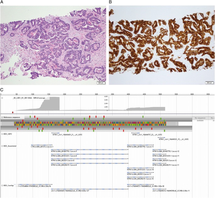
Figure 3.
Liver biopsy analysis. (A) Haemotoxylin and eosin (H&E): A core biopsy of the patient’s liver mass demonstrated a moderately differentiated adenocarcinoma, morphologically compatible with pancreatobiliary origin (H&E, 100× original magnification). (B) TrkA immunohistochemistry (IHC): Immunohistochemical staining for TrkA (NTRK1) demonstrated diffuse, strong cytoplasmic expression (TrkA IHC, clone EP1058Y, Abcam, Cambridge, UK, 100× original magnification). (C) Archer® software: Fusion analysis was carried out on the tumoral RNA with the MSK-IMPACT™ panel and demonstrated an in-frame fusion between CTRC (NM_007272) exon1 and NTRK1 (NM_002529) exon8, including the kinase domain of NTRK1 (JBrowse software).

