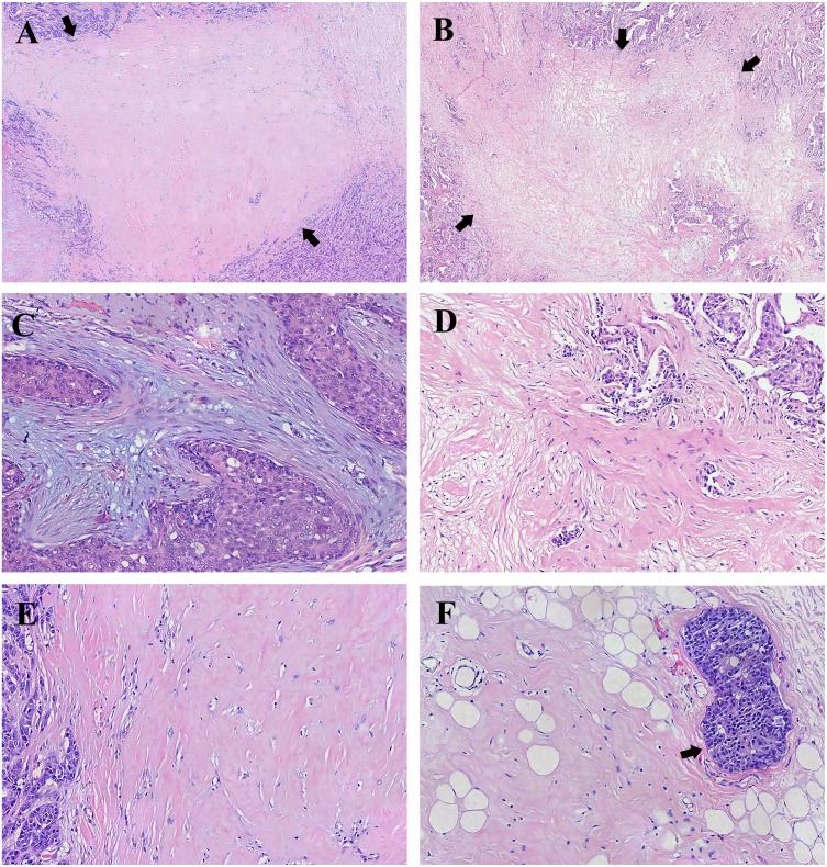
Figure 1. Representative histology of fibrotic focus (FF) in breast invasive ductal carcinoma (IDC).
(A) A representative histology of FF with the appearance of scar-like lesion indicated by arrows (H&E; magnification, ×20). (B) A representative histology of FF with the appearance of irregular moth-eaten radiating fibrosclerotic core indicated by arrows (H&E; magnification, ×20). (C) FF with mild fibrosis showing high number of fibroblasts and small amount of collagen fibers in stroma (H&E; magnification, ×100). (D) FF with moderate fibrosis intermediating between mild fibrosis and high fibrosis and numerous tumor nests in FF (H&E; magnification, ×100). (E) FF with high fibrosis showing mostly hyalinized collagen fibers (H&E; magnification, ×100). (F) High fibrosis and peripheral vascular invasion indicated by the arrow (H&E; magnification, ×100). Photograph credit: Doctor Xiangtao Lin.

