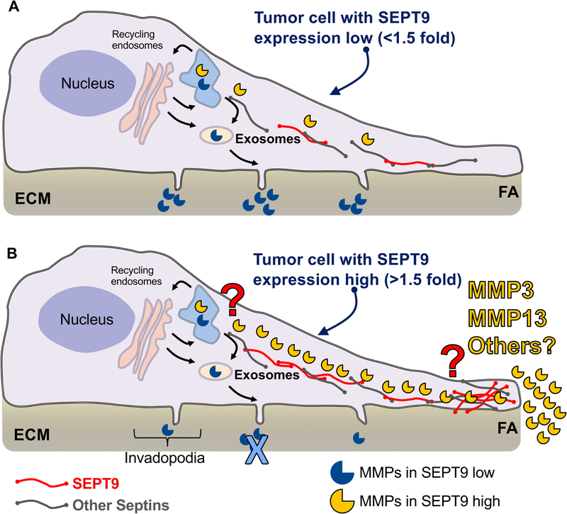
Figure 8: General model of functional consequences of SEPT9 overexpression in breast cancer cells.
A. In the absence of SEPT9 upregulation, degradation of the ECM in cancer cells occurs at invadopodia (protrusions that assemble at the ventral side of cells). After synthesis, MMPs - mainly MMP2 and MMP9 (open mouth blue circles) - are directed to the endoplasmic reticulum (ER, light blue structure at the top of the cell) and after Golgi processing (pink large vesicular structures), MMPs are delivered to the plasma membrane primarily via exosome secretion. B. In SEPT9-overexpressing cells, septin filaments (red = SEPT9 and gray = other septins) assemble into a network directed to FAs. The septin network promotes the delivery of MMP3 and MMP13 (open mouth yellow circles) to FAs, which represents the main ECM degradation pathway in these cells. The mechanisms by which SEPT9 and possibly other septins promote MMP3 and MMP13 secretion at FAs remains to be elucidated (red question mark). Likewise, whether SEPT9 promotes the secretion of MMPs or ECM-degrading enzymes other than MMP3 and MMP13 at FAs requires further investigation.

