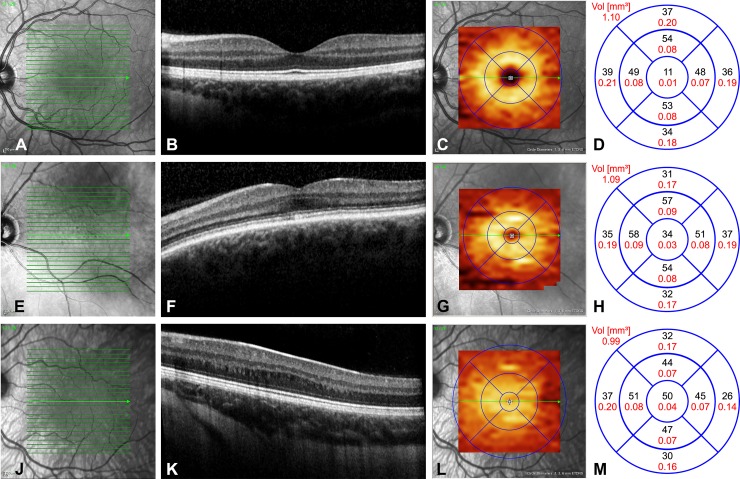
Fig 2.
Representative images of left eyes from the three patient groups: A-D healthy controls, E-H non-albinism FH, I-L albinism FH. In healthy controls (Fig 2A–2D), foveal pit formation is detectable through extrusion of the inner retinal layers, ONL widening and lengthening of photoreceptor outer segments (2B). The GCLT distribution pattern forms a doughnut shaped area around an inner annulus (2C). Values of GCLT in μm (black letters) and mm2 (red letters) in the nine areas are given in Fig 2D, showing a circular and homogenous GCLT distribution depending on the eccentricity of the foveal pit with RGC accumulation in the intermediate ring. In a patient with non-albinism FH grade 1 (Fig 2E–2H), the extrusion of the plexiform layers was absent, whereas the other foveal structural features were present (2F). The doughnut shape of the GCLT distribution was still recognizable (2G), although the central area showed markedly higher GCLT values (2H) due to shallower pit formation. In the albinism FH patient (Fig 2I–2L) the fovea could be located only through the widening of the ONL, representing FH grade 3 (2J). The GCLT was markedly reduced in area t II and shifted towards the centre where GCLT values were increased (2L).

