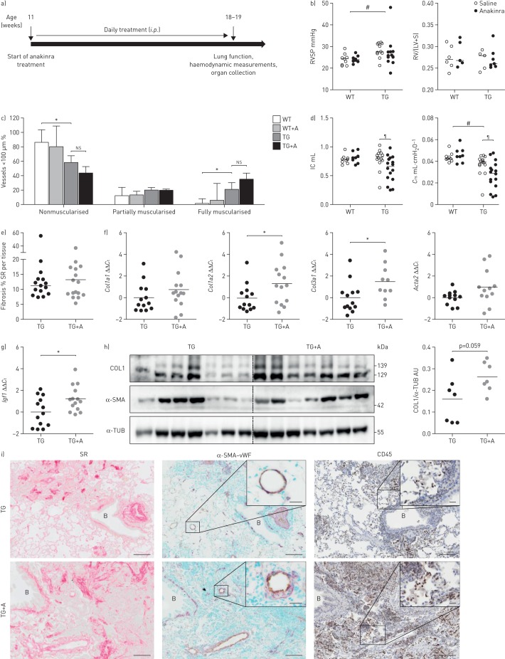
FIGURE 4.
Blocking of interleukin (IL)-1 signalling worsens lung function and increases extracellular matrix production in fos-related antigen-2 (Fra-2)-overexpressing (transgenic (TG)) mice. RSVP: right ventricular systolic pressure; RV: right ventricle, LV: left ventricle; S: septum; ns: nonsignificant; IC: inspiratory capacity; Crs: compliance of the respiratory system; SR: Sirius red; Ct: cycle threshold; COL1: collagen 1; α-SMA: α-smooth muscle actin; α-TUB: α-tubulin; AU: arbitrary units; vWF: von Willebrand Factor. a) Overview of anakinra treatment: Fra-2 TG mice received 25 mg·kg−1 anakinra per day as intraperitoneal injections for 8 weeks and were sacrificed at the age of 18–19 weeks. b) RVSP as determined by right heart catheterisation and the Fulton index (RV/(LV+S)) of Fra-2 TG and WT mice with anakinra treatment or vehicle control (saline). #: p<0.05, significance of genotype effect determined by two-way ANOVA. c) Percentage of nonmuscularised, partially muscularised and fully muscularised vessels <100 µm in diameter. n=3 for WT and n=9 for TG; mean±sd. *: p<0.05, unpaired t-test. d) Lung function measurements (IC and Crs) of Fra-2 TG and WT mice with anakinra treatment or vehicle control (saline). #: p<0.05, significance of genotype effect determined by two-way ANOVA; ¶: p<0.05, significance of the difference between Fra-2 TG with and without anakinra treatment determined by two-way ANOVA with Bonferroni's post-test. e) Morphometric quantification of collagen on SR-stained lung slides from Fra-2 TG and WT mice with (TG+A) and without (TG) anakinra treatment. f, g) Quantitative real-time PCR analysis of f) Col1a1, Cola1a2, Col3a1 and Acta2 and g) Igf1 expression in Fra-2 TG mice. ΔCt values were normalised to the mean of the untreated Fra-2 TG group (ΔΔCt). B2m (β2-microglobulin) and Hmbs (hydroxymethylbilane synthase) were used as reference genes. *: p<0.05, unpaired t-test. h) Western blot analysis and quantification of COL1 and α-SMA levels in Fra-2 TG and TG+A mice lung homogenates. α-TUB served as a loading control. i) Collagen staining with SR (collagen in red), immunohistochemical double staining of vessels against α-SMA (violet) and endothelium marker vWF (light brown), and immunohistochemical staining against inflammatory cell marker CD45. B: bronchi. Scale bar: 100 µm (main)/20 µm (insets).

