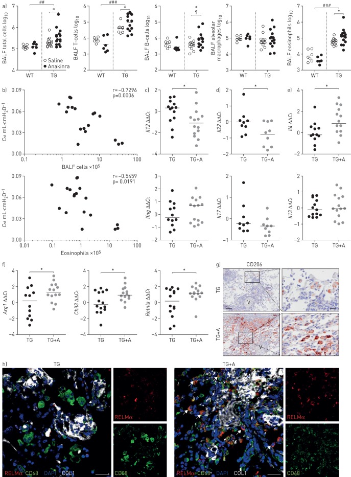
FIGURE 6.
Blocking of interleukin (IL)-1 signalling increases inflammation in fos-related antigen-2 (Fra-2)-overexpressing (transgenic (TG)) mice. BALF: bronchoalveolar lavage fluid; WT: wild-type; Cst: quasi-static lung compliance; Ct: cycle threshold; RELMα: resistin-like molecule-α; COL1: collagen 1; DAPI: 4′,6-diamidino-2-phenylindole; qRT: quantitative real-time. a) Flow cytometric analysis of inflammatory cell populations in the BALF of Fra-2 TG and WT mice with anakinra or vehicle control (saline) treatment. ##: p<0.01; ###: p<0.001, significance of genotype effect determined by two-way ANOVA; ¶: p<0.05, significance of difference between Fra-2 TG with and without anakinra treatment determined by two-way ANOVA with Bonferroni's post-test; *: p<0.05, unpaired t-test. b) Correlation plots of Cst with BALF total cell count or BALF eosinophils. c–e) Relative expression levels determined by qRT-PCR of key inflammatory mediators in lung homogenates of Fra-2 TG mice upon anakinra treatment. ΔCt values were normalised to the mean of the untreated Fra-2 TG group (ΔΔCt). B2m (β2-microglobulin) and Hmbs (hydroxymethylbilane synthase) were used as reference genes. *: p<0.05, unpaired t-test. f) qRT-PCR analysis of markers of alternative macrophage polarisation. ΔCt values were normalised to the mean of the untreated control group (ΔΔCt). *: p<0.05, unpaired t-test. g) Immunohistochemical staining of CD206 (brown) on lung sections from Fra-2 TG mice with (TG+A) and without (TG) anakinra treatment. V: vessel. Scale bar: 100 µm (main)/20 µm (insets). f) Immunofluorescence staining of RELMα (red), CD68 (green), COL1 (white) and DAPI (nuclei; blue). Arrows: CD68+/RELMα+ double-positive cells. Scale bar: 25 µm.

