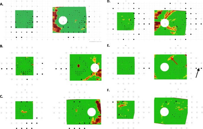
Figure 4.
Same as in Figure 3 for the six DG eyes that failed to show aS-aF agreement. (A–C) Three eyes with abnormal arcuate regions that were more obvious on the 24-2 VFs (A–C), and 10-2 VFs (A, B) probability maps than they were on the OCT probability maps. (D) An eye with an arcuate defect seen on 24-2 and on the OCT RNFL plot; however, the abnormal VF and OCT regions did not spatially overlap. (E, F) Two eyes with abnormal regions on both OCT and VFs that are subtle and/or difficult to discern.

