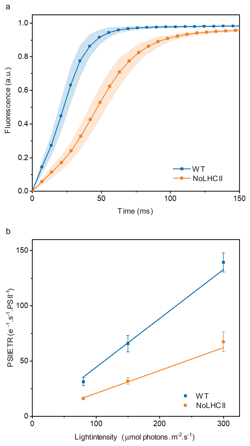
Figure 3.
Functional antenna size of PSII. (a) Chl fluorescence induction curves of both WT and NoLHCII measured in the presence of DCMU using sub-saturating light of 80 μmol photons m−2 s−1. (b) The light-limited, maximal PSII electron transport rate (ETR) of WT and NoLHCII determined using sub-saturating light of 80, 150 and 300 μmol photons m−2 s−1. See Supplementary Table 1 for the fitting parameters. The data in both panels represent the mean ± s.d. (n = 5 biologically independent experiments)

