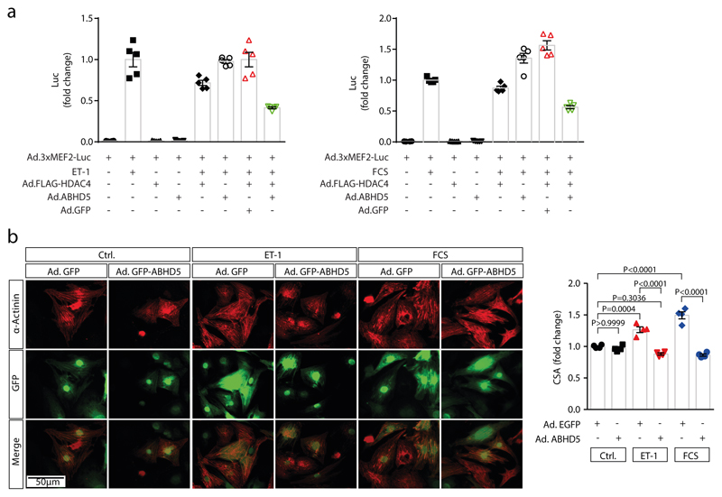
Extended Data Fig. 4. ABHD5 inhibits MEF2 and cardiomyocyte hypertrophy.
(a) NRVMs were transduced with the Ad3xMEF2C-Luc reporter. Ad.FLAG-HDAC4, Ad.ABHD5 plus GFP on a bicistronic promoter and Ad.GFP (10 MOI) were then co-expressed as indicated and NRVMs were stimulated for 24 hrs with endothelin-1 (ET-1) or fetal calf serum (FCS); n=5 (n represents independent samples) and values are presented as Mean±SEM. (b) Right panel: Fluorescence microscopy of NRVMs expressing Ad.ABHD5 plus GFP on a bicistronic promoter or Ad.GFP (10 MOI each). NRVM hypertrophy was induced with 100 nM ET-1 or 10% FCS for 24 hrs. Shown are representative images of α-actinin staining (red) of NRVMs and GFP fluorescence to confirm adenoviral expression. Images are representative of at least three independent experiments. Scale bar=50 μm. Right panel: Quantification of NRVM cross sectional areas (CSA). Statistical analysis: Values are presented as Mean±SEM, n=4 (n represents independent samples); by one-way ANOVA, P<0.05 considered as significant.

