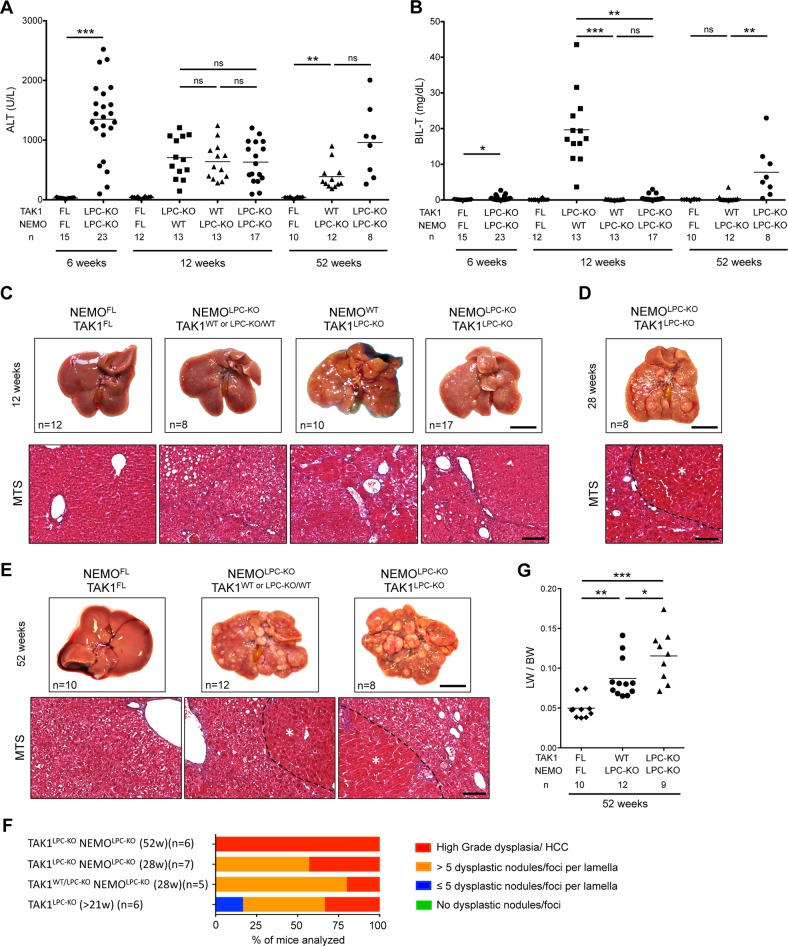
Fig. 6.
NEMO deletion strongly prevents biliary damage but leads to only a marginal delay in HCC progression in TAK1LPC-KO mice. a, b Serum levels of ALT (a) and total Bilirubin (b) in 6-, 12- and 52-week-old mice with the indicated genotypes. c–e Representative liver photos and images of liver sections stained for Masson’s trichrome from 12- (c), 28- (d), and 52-week-old mice (e) with the indicated genotypes. Dysplastic nodules or HCCs are outlined and marked with an asterisk in d and e. f Histopathological evaluation of hepatocarcinogenesis in liver samples from mice with the indicated age. g LW/BW ratio in 52-week-old mice with the indicated genotypes. The number of mice analyzed (n) is indicated in every graph. All graphs show mean values of the individual data points. *p < 0.05, **p < 0.01, ***p < 0.005. Bars: c, e upper, 1 cm; lower, 100 μm

