
Figure 3.
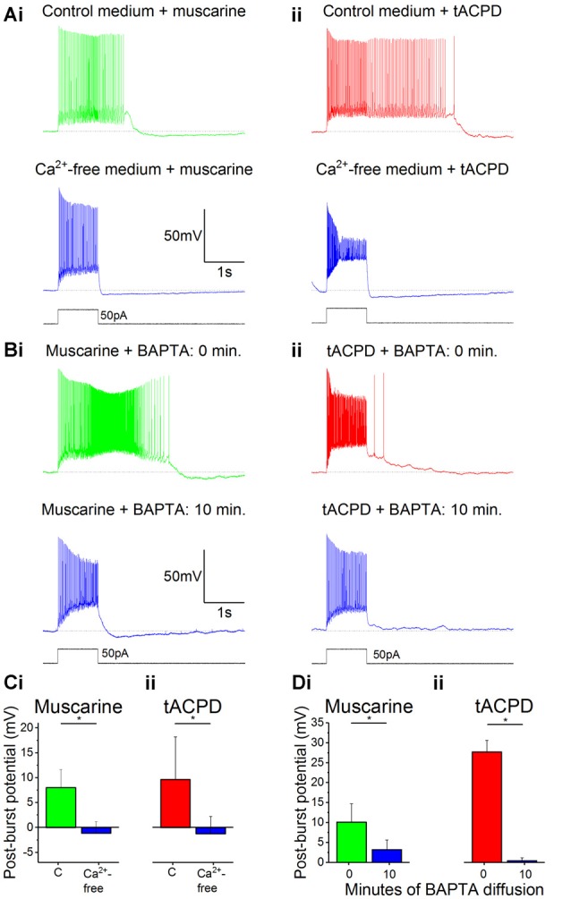
The muscarinic and glutamatergic plateau potentials are calcium-dependent. (A) Example traces of the abolition of muscarine- (Ai) or tACPD-induced (Aii) plateau potentials by switching to a Ca2+-free artificial cerebrospinal fluid (aCSF) medium in the continuous presence of agonists. (B) Example traces showing the abolition of muscarine- (Bi) or tACPD-induced (Bii) plateau potentials immediately after (upper traces) and 10 min after break-in (lower traces) when the BAPTA-containing pipette solution became contiguous with the intracellular space. (C) Summary plots of the change in post-burst potential in the presence of either muscarine (Ci; n = 6) or tACPD (Cii; n = 7) after switching to a Ca2+-free medium, showing a move from an ADP to an AHP. (D) Summary plots of the change in post-burst potential in the presence of muscarine (Di; n = 5) or tACPD (Dii; n = 8) after 10 min of allowing BAPTA to diffuse into the intracellular solution. Error bars = SEM. *P < 0.05.