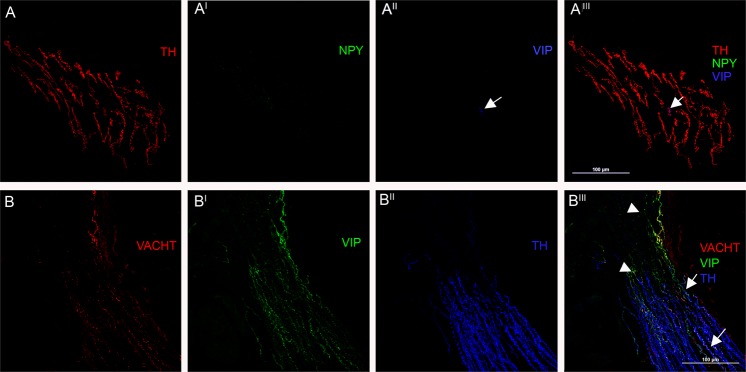
Figure 2.
Autonomic nerves in Arrector pili muscles. Confocal microscope analysis (x400) of arrector pili muscle. (A) Nearly all of these fibers are adrenergic showing a staining against TH (red). Interestingly these fibers did not express NPY (green), which is different from the adrenergic fibers innervating arterioles that co-expressed NPY. This arrector pili muscle also shows a VIP positive fiber (arrows) in blue. This fiber co-expressed TH and represents a sympathetic cholinergic fiber. (B) Different sympathetic cholinergic fibers co-expressing TH (blue), VIP (green) and VACHT (red) staining can be seen in this arrector pili muscle (arrows). A few VIP fibers did not express TH and VACHT (head of arrows) and they are likely to be somatic fibers.

