Figure 2.
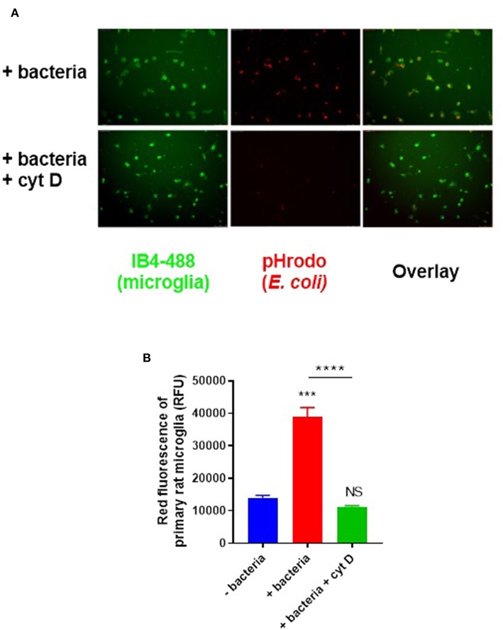
Primary rat microglia rapidly phagocytose E. coli bacteria in vitro. (A) Primary rat microglia phagocytose pHrodo-conjugated E. coli over 60 min in culture, which was prevented by cytochalasin D (10 μM). (B) Microglial phagocytosis of pHrodo-conjugated E. coli was quantified as mean red fluorescence via flow cytometry; there is a significant increase in microglial fluorescence after 60-min with E. coli which is abolished by cytochalasin D, when compared to the “-bacteria” control. Values are means ± SEM of at least three independent experiments. Statistical comparisons were made via one-way ANOVA. NS p ≥ 0.05, ***p < 0.001, ****p < 0.0001 vs. controls, except where indicated by bars over relevant columns.

