
Fig. 13.
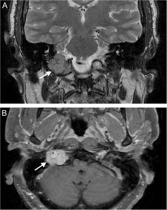
Jugular paraganglioma. A 55-year-old woman with relatively sudden onset hoarseness and dysphagia to liquids, who was found to have right vocal cord paralysis on laryngoscopy. Coronal T2 (a) and axial T1 post-contrast (b) MR images reveal a T2-isointense, avidly-enhancing lesion at the right jugular fossa (arrows) extending intracranially, with mild mass effect upon the right cerebellar hemisphere. Foci of low signal (“pepper”) represent flow voids, reflecting hypervascularity. Based on location and appearance, findings are consistent with a jugular paraganglioma (glomus jugulare tumor)