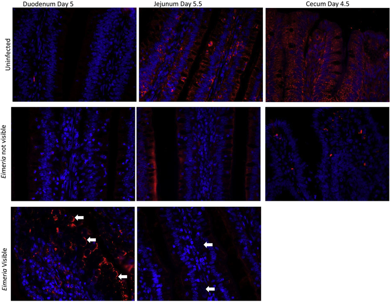
Figure 4: Interferon Gamma Immunofluorescent Histochemistry Images.
IFNγ presence is quantified using image J to calculate the percent staining of the imaged intestinal section. Chicks were orally gavaged with a 10X dose of an avirulent coccidia vaccine that contained E. acervulina, E. maxima, and E. tenella. Images shown are at time points in each section of the intestine when the largest difference of IFNγ presence was evident. Immunohistochemical staining of IFNγ protein (red) and cellular nuclei (blue) in control (top), null (middle) and infected (bottom) chicken mucosa. White arrows indicate areas where Eimeria are present. The intestinal lumen is located at the top of each photo. On day 4.5, identifiable Eimeria stages were not visualized in the cecum, therefore only uninfected and null images are shown.

