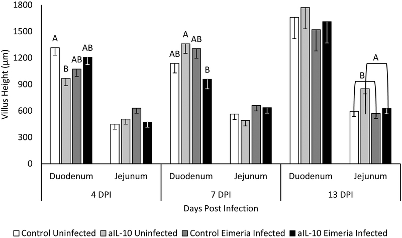
Figure 6: Villus Height.
Chickens were infected with Eimeria at 3 days of age and samples were taken at 4, 7 and 13 DPI. Villus height is measured from tip of villi to start of crypt in micrometers. Cecal measurements are not included because by definition, the ceca lack villi. Each bar represents the mean villus height of 3 chicks. Error bars indicate SEM. A,B Indicates significant difference, P<0.05 for each intestinal section and day post infection.

