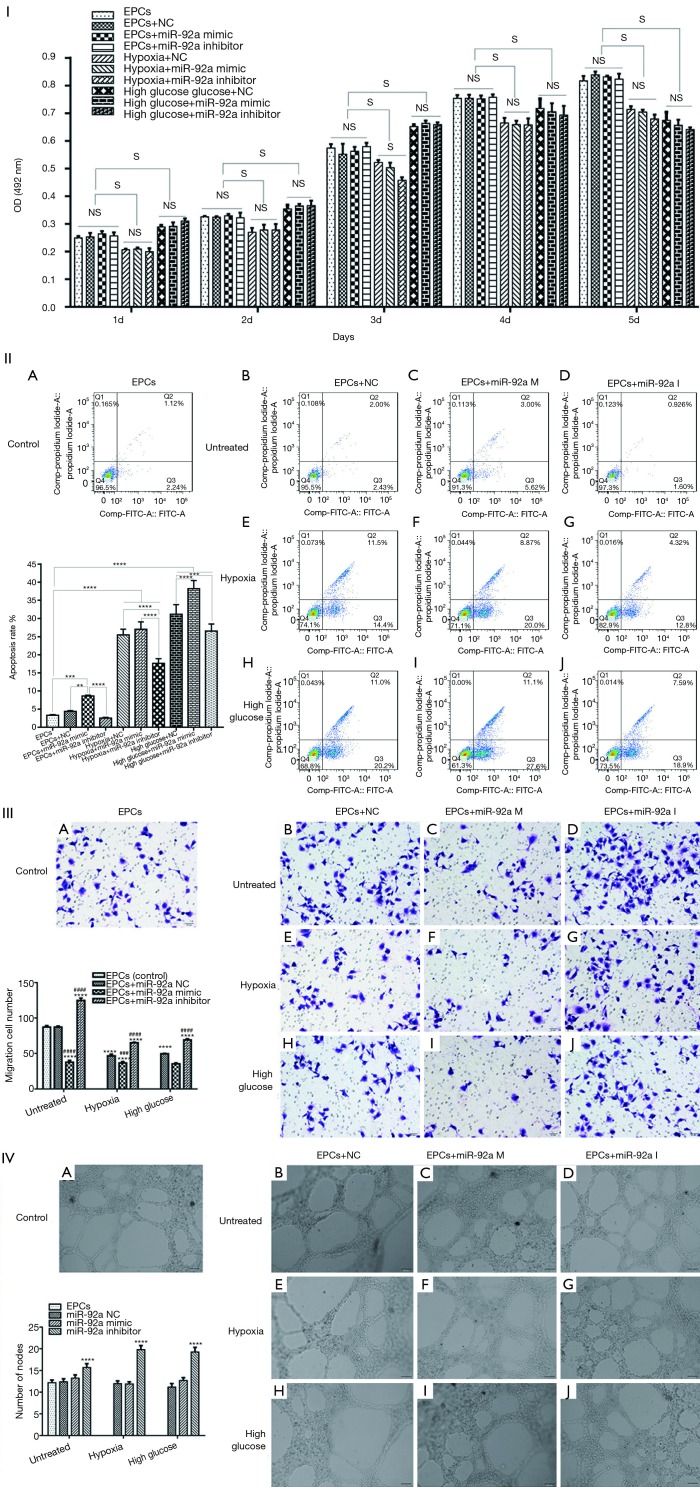
Figure 2.
Cell proliferation, apoptosis, migration, tube formation after various treatments EPCs underwent miR-92a inhibition or miR-92a over-expression under the condition of HO or HG. I: MTT assay was applied to detected proliferation of EPCs (S: significant difference; NS: no significant difference). II: Cells were harvested for the detection of apoptotic cells by flow cytometry (***, P<0.001, ****P<0.0001). III: migration ability of EPCs was tested by transwell assay (****, P<0.0001 vs. control group; ###, P<0.001, ####, P<0.0001 vs. miR-92a NC group) (0.1% crystal violet, 200×). IV: angiogenesis was evaluated by tube formation test (****, P<0.0001 vs. control group and miR-92a NC group).

