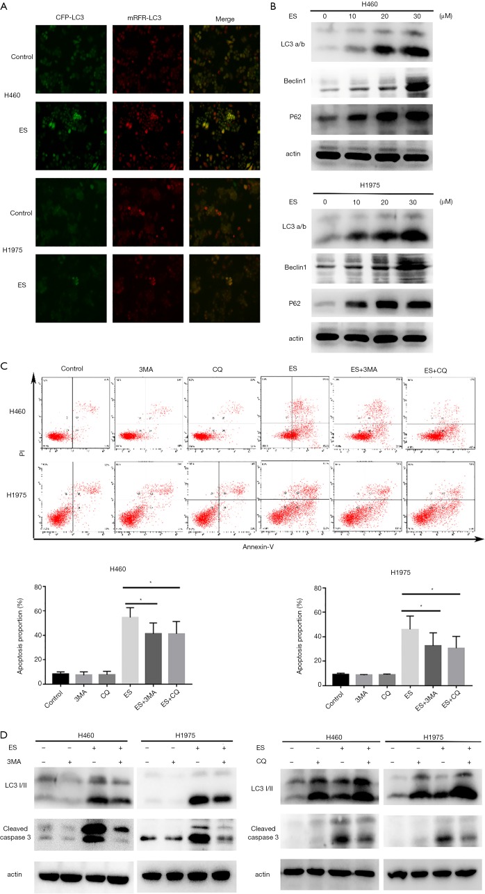
Figure 3.
ES induces autophagy, which contributes to cell death. (A) Images of mRFP-GFP-LC3 adenovirus infection monitored the autophagy flux in H460 and H1975 cells under fluorescence microscope (200× magnification); (B) the expression levels of indicated proteins were analyzed by western blot assay; (C) pretreated with 3-MA (5 mmol/L) or CQ (20 µmol/L) for 1 h, apoptosis was analyzed by FACS using an Annexin V-FITC/PI cell apoptosis kit; (D) cells were pretreated with 3-MA (5 mmol/L) or CQ (20 µmol/L) for 1 h. Expression levels of indicated proteins were detected by western blot assay. All data are expressed as mean ± SD. *, P <0.05; **, P<0.01; ***, P<0.001 compared to the control group. ES, Ecliptasaponin A; 3-MA, 3-methyladenine; CQ, chloroquine; FACS, fluorescence-activated cell sorting; PI, propidium iodide.

