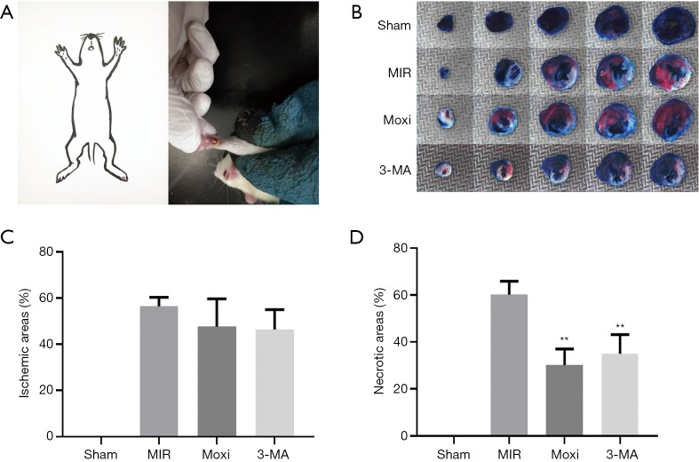
Figure 1.
Moxibustion preconditioning decreased necrotic areas of MIRI (n=6 each group). (A) The location of PC6 and moxibustion intervention; (B) representative photographs of ventricular tissue sections with Evans blue-TTC staining, showing 3 main zones: infarction (white), at-risk for infarction (red), and no infarction (blue); (C,D) averaged percentages of ischemic and necrotic areas, respectively. There was no significant difference in the ischemic area among the 3 groups, but the necrotic area was significantly smaller in the 3-MA and Moxi groups compared to the MIR group. All data are expressed as mean ± SD. **, P<0.01 vs. MIR group. MIRI, myocardial ischemia reperfusion injury.

