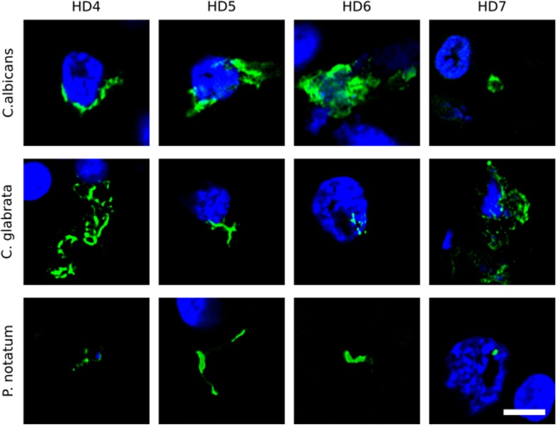
FIGURE 1.
Immunohistochemistry of striatum sections from Huntington’s disease patients using a battery of antifungal antibodies. The striatum CNS region of four HD patients (HD4–HD7) was processed for immunohistochemistry as described in section “Materials and Methods.” Paraffin sections were immunostained with rabbit polyclonal antibodies against C. albicans, C. glabrata, and P. notatum (green). Nuclei were stained with DAPI (blue). Scale bar: 5 μm.

