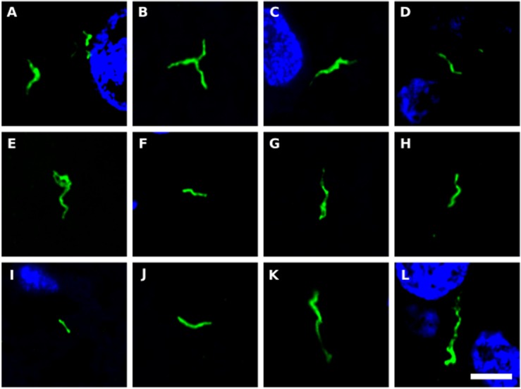
FIGURE 8.
Prokaryotic structures in Huntington’s disease brain sections using an anti-C. pneumoniae antibody. Different sections from HD patients were incubated with a rabbit polyclonal antibody against C. pneumoniae (green). DAPI staining is shown in blue. (A,C,E,F,H,J,K) striatum region; (B,D,G,I,L) frontal cortex region; (A,B) HD1; (C,D) HD2; (E) HD3; (F,G) HD4; (H,I) HD5; (J) HD6; (K,L) HD7. Scale bar: 5 μm.

