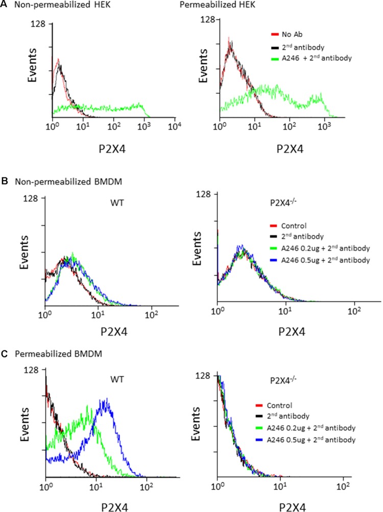
Figure 3.
Flow cytometry analysis of mouse P2X4 expression using Nodu 246. (A) Cell surface and intracellular expression of mouse P2X4 by P2X4-transfected and non-transfected HEK cells was assessed by flow cytometry after sequential incubation of cells with rat mAb Nodu 246 followed by a secondary PE-conjugated anti-rat-IgG antibody (2nd antibody), in non-permeabilized or permeabilized cells. Similar experiments were performed with BMDM obtained from WT and P2X4−/− mice. Cells were either non-permeabilized (B) or permeabilized (C). Note that permeabilization greatly enhanced the detection of mouse P2X4 in BMDM, further supporting the intracellular localization of the native receptor.

