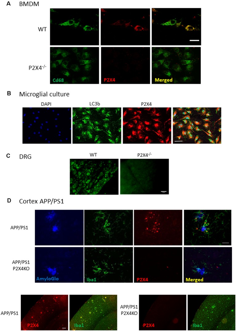
Figure 4.
Immunocyto- and -histochemistry with Nodu 246. (A) Co-immunostaining of WT and P2X4−/− BMDM with Nodu 246 and the lysosomal marker CD68. Scale bar: 5 μm. (B) Co-immunostaining of WT and P2X4−/− cultured mouse microglia using the rat-monoclonal antibody Nodu 246 and the lysosomal marker LC3b. Scale bar: 10 μm. (C) Immunostaining of dorsal root ganglion (DRG) of WT and P2X4−/− DRG. Scale bar: 50 μm. (D) Top panel, co-immunostaining of microglial cells (green, Iba1) and P2X4 (red, Nodu 246) and amyloid plaques (blue, AmyloGlo) in the cortex of brains from APP/PS1 and APP/PS1; P2X4−/− mice. P2X4 is localized in the intracellular compartments of activated microglia clustered around amyloid deposits. Scale bar 20 μm. Bottom panel, low magnification of the cortical region of APP/PS1 and APP/PS1; P2X4−/− mice stained with anti-Iba1 (green) and Nodu 246 (red). Note the dim P2X4 immunostaining in cortical neurons. Scale bar 50 μm.

