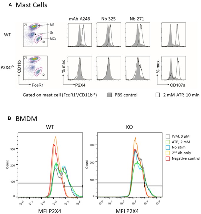
Figure 6.
Flow cytometry analysis of native mouse P2X4 expressed by peritoneal mast cells and BMDM. (A) Peritoneal cells from WT and P2X4−/− were stimulated (white) or not (gray) for 10 min with 2 mM ATP at 37°C. Cells were washed and incubated at 4°C with anti-CD11b-PerCO and anti-FcεR1-PE as well as with various antibodies directed against P2X4 and appropriate secondary antibodies (AF647 or AF488). CD107a (LAMP1-FITC) was used as a control experiment. Mast cells were identified as CD11b low, FcεR1+. mAb Nodu 246: rat monoclonal antibody; Nb 325, Nb 271: Nanobodies. (B) Similar flow cytometry experiments of WT and P2X4−/− BMDM using Nb 271 RbhcAb. Cells were stimulated as above with 2 mM ATP (ATP, green), 3 μM ivermectin (IVM, gray), or not stimulated (nanobody, blue). Negative controls were secondary antibody only (secondary antibody, orange) and unlabeled cells (Neg, red).

