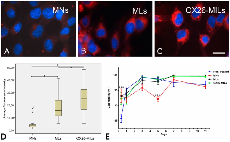
Figure 3.
The uptake of red fluorescent nanoparticles in RBE4 cells, and cell viability. Cells were incubated with 1 µg of magnetic nanoparticles (MNs), magnetic liposomes (MLs), or OX26-immunomagnetic liposomes (OX26-MLs). (A) Uptake is virtually absent in cells incubated with magnetic nanoparticles (MNs) without a liposome coat. Magnetic liposomes (MLs) (B) and magnetic OX26 immunoliposomes (OX26-MLs) (C) exhibit prominent fluorescence inside cells, indicative of uptake. Scale bar = 10 µm. (D) Quantitative estimate of the fluorescence emitted by RBE4 cells after the addition of magnetic nanoparticles (MNs), magnetic liposomes (MLs), or magnetic OX26 immunoliposomes (OX26-MILs) based on computed mean fluorescence intensity of 30–50 individual measurements in each group. The three groups are all significantly different (p ≤ 0.05), with MNs displaying the lowest intensity and OX26-MLs the highest. Outlier values are marked with asterisks numbered 4 and 5, due to their falling outside the definition as being values between 1.5 and three box lengths from the upper and lower edge of the box, respectively. (E) Cell viability of non-treated RBE4 cells or RBE4 cells treated for 8 h of incubation with MNs, MLs, and OX26-MILs. After 8 h, the viability of non-treated cells was significantly lower (p ≤ 0.001) than all the treated cells. At day five, the viability of MNs was significantly lower (p ≤ 0.001) than the non-treated cells and cells with MLs and OX26-MILs.

