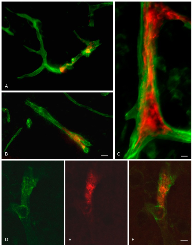
Figure 4.
Brain distribution of magnetic OX26 immunoliposomes following in situ perfusion without application of external magnetic force. Images are from rats which received 10 µg/g bodyweight of magnetic OX26 immunoliposomes. (A–C) Magnetic OX26 immunoliposomes (red) present inside brain capillaries do not distribute past laminin (green) which marks the basal membrane of brain capillaries. (D–F) Co-detection of laminin (green) and magnetic OX26 immunoliposomes (red) using confocal imaging. The pictures show a Z-stack generated from serial analyses of a single brain capillary. (F) Merging the two fluorophores reveals that magnetic OX26 immunoliposomes are confined to the interior of the brain capillary. Scale bars = 20 µm (A,B), 10 µm (C), and 5 µm (D–F).

