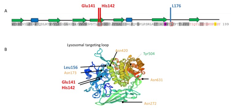
Figure 5.
Localization of GUSB mutations. (A), Figure adapted from Hassan et al [12,13]. Amino acid sequence of human GUSB centered on the substitutions (in red boxes) carried by the present case. Residues shaded in dark and light grey correspond to degree of conservation between species. Residue highlighted in pink is a glycosylation site. On the top of sequence, in the secondary structure of GUSB, green arrows indicate β-strands and blue boxes correspond to α-helices. (B), Three-dimensional structure of GUSB monomer (https://swissmodel.expasy.org/). In orange, glycosylation sites; in green, active-site residue; in red and blue, the maternally (c.422A>C;424C>T) and paternally (c.526C>T) inherited mutated residues in the patient, respectively.

