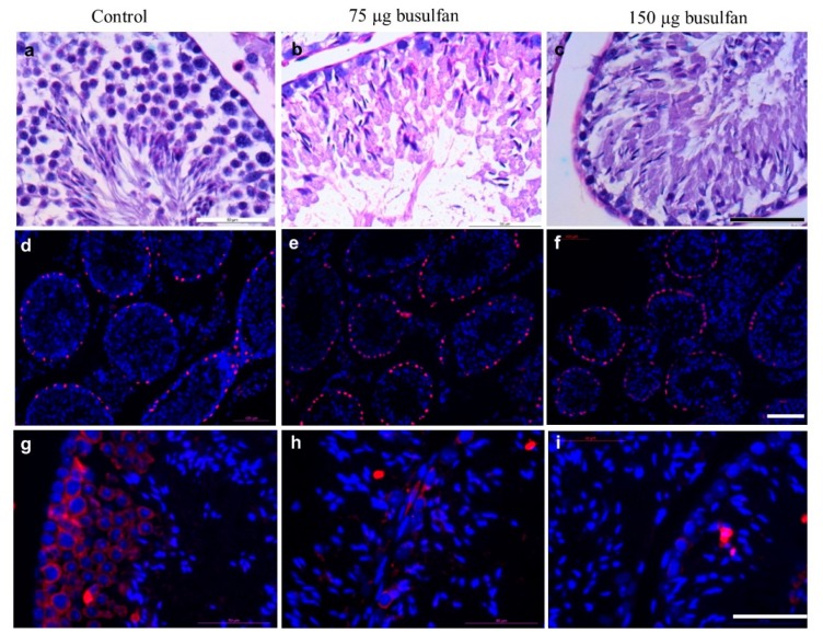
Figure 6.
Testis from nude mice sterilized by intratesticular busulfan injection. a–c: Cross sectional histological analysis of mouse testis by periodic acid–Schiff (PAS) staining. (a) nontreated control (b) two weeks after intratesticular busulfan injection with either 75 µg or (c) 150 µg. Scale bar: 50 µm. d–f: Cross sectional immunofluorescent analysis of mouse testis by Sertoli cell marker SRY-Box 9 (SOX9; red) and DNA visualization by DAPI staining (blue) (d) non-treated control (e) two weeks after intratesticular busulfan injection with either 75 µg or (f) 150 µg. Scale bar: 100 µm. g–i: Cross sectional immunofluorescent analysis of mouse testis by germ cell marker VASA (red) and DNA visualization by DAPI staining (blue) (g) non-treated control (h) two weeks after intratesticular busulfan injection with either 75 µg or (i) 150 µg. Scale bar: 50 µm.

