Figure 7.
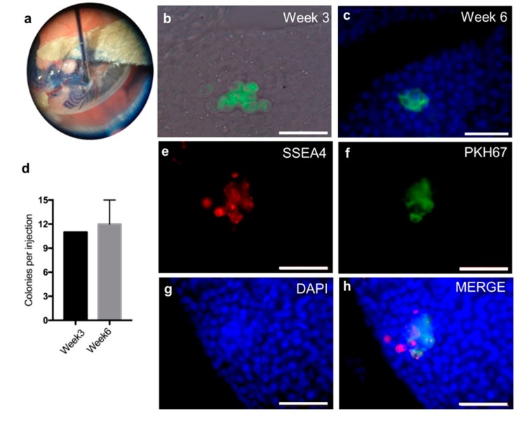
Xenotransplantation of the human cells into busulfan treated nude mice. (a) Demonstration of successful transplantation of human SSC-like cells into murine seminiferous tubules using trypan blue. (b) PKH67 (a green fluorescent cell linker) pre-labelled cells forming colonies in recipient testis merged with bright channel three weeks after transplantation. (c) PKH67 pre-labelled cells forming colonies in recipient testis merged with DAPI staining six weeks after transplantation. (d) Mean number with standard deviation of colonies formed numbers in recipient mouse testis. Three weeks after transplantation (n = 1) and six weeks after transplantation (n = 3). e–h: Whole mount immunofluorescent analysis of mouse testis six weeks after transplantation by SSC marker, stage-specific embryonic antigen-4 (SSEA4; red) and DNA visualization by DAPI staining (blue) (e) SSEA4; (f) PKH67; (g) DAPI and (h) merged pictures from previous three channels. Scale bar: 50 µm.

