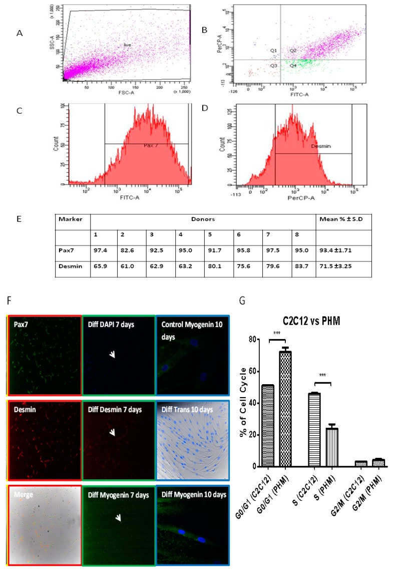
Figure 7.
The phenotypic characterization of primary human myoblasts. (A–D) Flow cytometric analysis of Primary Human myoblasts (PHMs) was performed as described in Materials and Methods section. Average of cells (%) staining positive for Pax7 and desmin are shown. (E) Several preparations of the PHMs were analyzed and shown is representative of eight donors. Results are shown as mean ± S.D. (F) Red outlined column (first column): Preliminary characterization of isolated cells as performed to ascertain whether the culture generated contained myoblasts: Pax7 (green), desmin (red) (fluorescent microscopy at 100× magnification). Green outlined Column (second column): PHMs were exposed to differentiation inducing conditions for 7 days and stained via immunofluorescence for desmin (red), myogenin (green) and nuclei (blue). White arrows indicate multinucleated myofibres. Blue outlined Column (third column): PHMs were able to preserve their differentiation ability after long periods of sub-culturing. Cells were maintained and passaged for two months (imaged by confocal microscopy (myogenin) at 400× magnification or fluorescent microscopy (Trans) at 200× magnification). (G) Comparison of C2C12 and PHMs. Cells were analyzed through flow cytometry and percentages of cells in each cell cycle stage quantified. * p < 0.05; ** p < 0.01; *** p < 0.001.

