Figure 2.
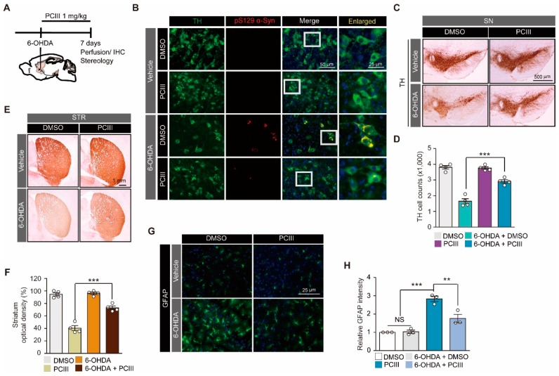
PCIII prevents 6-OHDA-induced α-synuclein aggregation and dopamine cell death in vivo. (A) The schematic experimental schedule of PCIII therapeutic evaluation in 6-OHDA (8 µg) induced PD mouse models. IHC, immunohistochemistry. To model the dopaminergic neuronal death in mice, 6-OHDA was stereotaxically injected into the striatum (coordinates from bregma, L: -2.0, AP: 0.5, DV: -3.0 mm) of 2-month-old mouse brains. (B) Representative immunofluorescence images of TH (green) and serine 129-phosphorylated α-synuclein (pS129 α-Syn, red) expression in the ventral midbrain sections from 6-OHDA-induced PD mice treated with PCIII (i.p. 1mg/kg/day) or DMSO for seven days. (C) Representative tyrosine hydroxylase (TH) immunohistochemical staining of the substantia nigra of the 6-OHDA PD mice treated with PCIII (i.p. 1 mg/kg/day, 7 days) or DMSO. (D) Stereological assessment of tyrosine hydroxylase (TH)-positive dopaminergic neurons in the substantia nigra pars compacta of the injection side of the indicated mouse groups (n = 4 mice per group). (E) Representative tyrosine hydroxylase (TH) immunohistochemical staining of the striatum (STR) of 6-OHDA PD mice treated with PCIII (i.p. 1 mg/kg/day, 7 days) or DMSO. (F) Quantification of relative TH stained optical dopaminergic nerve terminal fiber densities in the striatum of the indicated experimental groups (n = 4 mice per group). (G) Representative immunofluorescence images of GFAP in the substantia nigra of the indicated mouse groups. (H) Quantification of relative GFAP fluorescence intensities in the substantia nigra sections from the indicated experimental groups (n = 3 mice per group). The quantified data are expressed as means ± SEMs. Statistical significance was determined by the ANOVA test with Tukey post-hoc analysis, ** p < 0.01, and *** p < 0.001. NS, nonsignificant.

