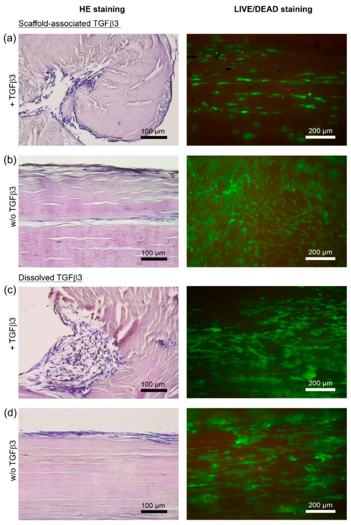
Figure 2.
Microscopic appearance of MSC-seeded tendon scaffolds treated with TGFβ3 (+TGFβ3) and the respective internal control scaffolds (w/o TGFβ3). Representative images of hematoxylin- and eosin-stained paraffin sections of MSC-seeded tendon scaffolds (a–d, left) and of corresponding LIVE/DEAD®-stained MSC-seeded tendon scaffolds (a–d, right). The panel of LIVE/DEAD®-stained scaffolds shows vital cells in green and cells with defect cellular membranes in red. MSC-seeded tendon scaffolds were either directly loaded with TGFβ3 (scaffold-associated) (a), or TGFβ3 was applied as a standard cell culture medium supplement (dissolved) (c). Respective internal control scaffolds—(b) internal control for scaffolds directly loaded with TGFβ3; (d) internal control for scaffolds that received TGFβ3 as a standard cell culture medium supplement—were not treated with TGFβ3 (w/o TGFβ3). Note the obvious alterations of the scaffold morphology (left panel of hematoxylin- and eosin-stained sections), illustrating the increased cell-mediated scaffold contractions in the presence of TGFβ3 regardless of the route of application ((a) directly applied TGFβ3 and (c) TGFβ3 applied as a cell culture medium supplement). All images shown were taken after 5 days from tendon scaffolds that were seeded with MSC from the same donor horse.

