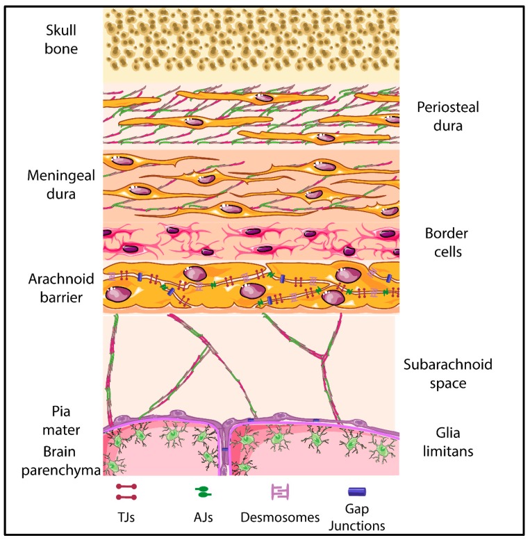
Figure 4.
Schematic representation of the meningeal layers. The different layers and cellular composition of the meningeal layers are displayed. Tight junctions are highlighted between the arachnoid barrier cells as parallel lines, with adherens junctions, desmosomes and gap junctions also being represented. The shapes of the cell types were adapted from Servier Medical Art (http://smart.servier.com/), licensed under a Creative Common Attribution 3.0 Generic License.

