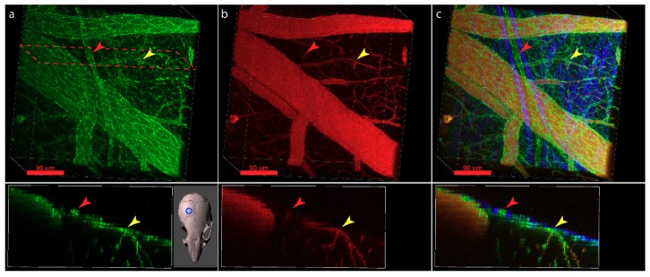
Figure 6.
Visualization of CNS and meningeal endothelial AJs using the VE-Cadherin-GFP reporter mouse. (a) Cranial window allowing to visualize meningeal, subarachnoid, subpial and cortical vascular VE-cadherin-GFP+ AJs. (b) Cranial window preparation from (a) highlighting blood vessels after i.v. injection of TRITC Dextran. (c) Overlay of a and b and the second harmonic generation of the collagen fibers in the dura allow to distinguish VE-cadherin-GFP+ TRITC+ blood vessels and VE-cadherin-GFP+ TRITCneg lymphatic vessels in the dura mater. Examples of blood and lymphatic vessels of similar caliber size are highlighted with a yellow and red arrowhead, respectively. The cranial window was placed over the right hemisphere of the mouse brain as depicted in the insert below (a). Top row—3D stack, bottom—XZ maximum intensity projection of 20 µm along Y at the cross section highlighted at the top. Scale bars = 90µm.

