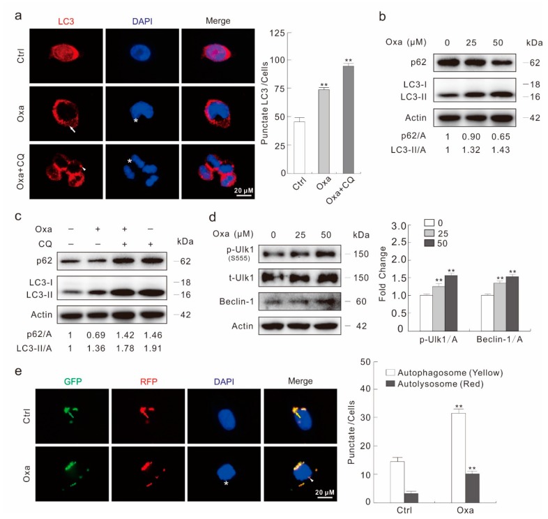
Figure 2.
Oxa induces autophagy in SW480 cells. (a) Immunofluorescence using the antibody of LC3 was performed in SW480 cells following treatment with Oxa (25 μM hereafter, or otherwise indicated) in the presence or absence of CQ (20 μM hereafter) for 2 h (1000 magnification). The numbers of the punctate LC3 in each cell were counted, and at least 30 cells were included for each group. (b–d) Cells were treated with indicated dose of Oxa for 2 h in the presence or absence of CQ. Cell lysates were subjected to immunoblotting with the antibodies indicated. (e) After transfection with GFP-RFP-LC3 plasmids for 24 h and split onto coverslips then cultured overnight, SW480 cells were treated with or without Oxa for 2 h (1000 magnification). The number of the yellow and red dots in each cell was counted, and at least 20 cells were included for each group. Data represent three independent experiments. ** p < 0.01 vs. control.

