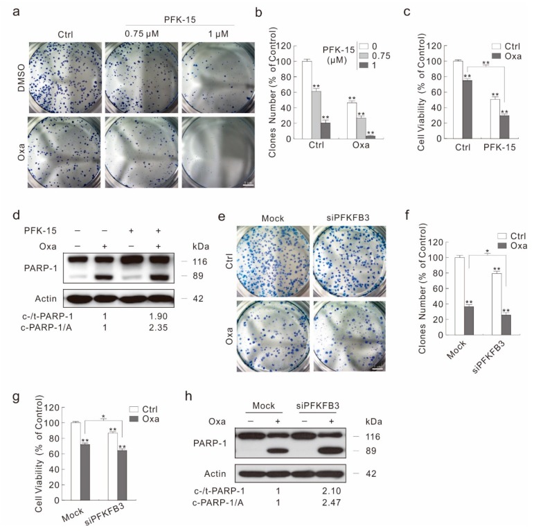
Figure 5.
PFKFB3 inhibition enhances the cytotoxicity of Oxa. (a,b) Colony growth assay was performed with Oxa (1.5 μM) in the presence or absence of PFK-15. (c,d) Cell viability was analyzed by MTS assay following the treatment with Oxa in the presence or absence of PFK-15 for 24 h (c); cell lysates from cells treated same as (c) were subjected to immunoblotting with indicated antibodies (d). (e–h) SW480 cells were transfected with the PFKFB3 siRNA or control siRNA for 48 h. Colony growth assay was performed with Oxa (1.5 μM) (e), and relative clones number to control was shown in (f). Cell viability was analyzed by MTS assay following the treatment with Oxa for 24 h (g). Cell lysates were subjected to immunoblotting with indicated antibodies following treatment with Oxa for 24 h (h). For histogram graph data, * p < 0.05 vs. control, and ** p < 0.01 vs. control. Similar experiments were repeated three times.

