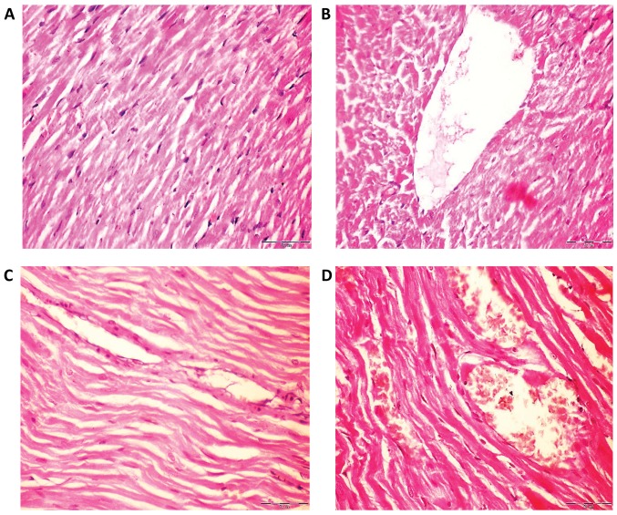
Figure 4.
Histological analysis of haematoxylin and eosin stained rat heart tissues of control, Fe2O3NP-exposed group, AgNP-exposed group, and Fe2O3NP and AgNP-coexposed group. (A) Control rats demonstrated normal myofiber cells and normal architecture. (B) Heart tissues of Fe2O3NP-exposed group revealed myocardial degeneration changes. (C) Heart of AgNP-exposed group revealed fragmentation of sacroplasm and degeneration changes in myocardial fibers. (D) Heart of the Fe2O3NP and AgNP-coexposed group revealed loss of cross striation and severe myocardial degeneration changes (magnification, ×400). Fe2O3NP, iron oxide nanoparticle; AgNP, silver nanoparticle.

