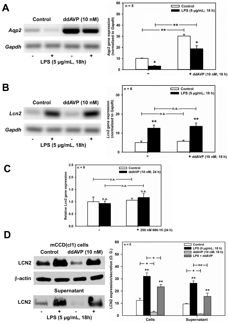
Figure 6.
ddAVP decreases Lcn2/LCN2 expression through a CREB-independent, posttranslational mode of action in mCCD(cl.1) cells. (A,B) Expression levels of Aqp2 (A) and Lcn2 (B) mRNA by RT-PCR in mCCD(cl.1) cells treated with ddAVP ± LPS. Data are means ± SEM of 5–6 experiments. Statistical analysis compares the expression levels of Aqp2 or Lcn2 in controls and ddAVP-treated cells ± LPS, by one-way ANOVA. n.s. = not significant. (C) Expression levels of Lcn2 mRNA by qPCR in mCCD(cl.1) cells treated with ddAVP ± 666-15. Data are means ± SEM of nine experiments. Statistical analysis compares relative expression levels of Lcn2 in controls and ddAVP-treated cells ± 666-15, by one-way ANOVA. n.s. = not significant. (D) Expression and secretion of LCN2 in mCCD(cl.1) cells treated with LPS, in the absence or presence of ddAVP. Media (supernatant), treated as described in Figure 5E, were immunoblotted. Cellular LCN2 protein expression was normalized to β-actin. Data are means ± SEM of five experiments. Statistical analysis compares expression levels of cellular and secreted LCN2 in controls and ddAVP-treated cells ± LPS, by one-way ANOVA. (E) Expression of LCN2 in mCCD(cl.1) cells treated with ddAVP for 6 h ± cycloheximide, a blocker of translational elongation, was detected by immunofluorescence microscopy of permeabilized cells. Hoechst 33342 counterstains nuclei. Statistical analysis shows means ± SEM of four experiments and comparison of the conditions ± ddAVP by unpaired t-test. n.s. = not significant. For a definition of asterisks, see “statistics” in the Methods.


