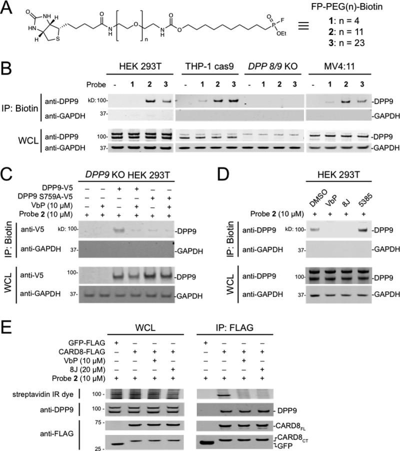
Figure 3.
An extended activity-based probe does not disrupt the DPP9–CARD8 interaction. (A) Structure of FP-PEG(n)-Biotin probes. (B–D) Streptavidin enrichment of the indicated cell lysates with the indicated FP-biotin probes. In C, lysates from DPP9 KO HEK 293T cells transiently transfected with constructs encoding the indicated DPP9 protein (1 μg) were treated with VbP or DMSO for 1 h. In D, lysates from HEK 293T cells were treated with VbP (10 μM), 8J (20 μM), or 5385 (20 μM) for 1 h. (E) Lysates from HEK 293T cells transiently transfected with constructs encoding the indicated FLAG proteins were treated with VbP or 8J for 1 h, followed by probe 2 for 1 h, and subjected to anti-FLAG IP. Immunoblots depict input whole cell lysate (WCL) and captured proteins from either FP-biotin enrichment (IP: Biotin) or anti-FLAG IPs (IP: FLAG).

