
Figure 1.
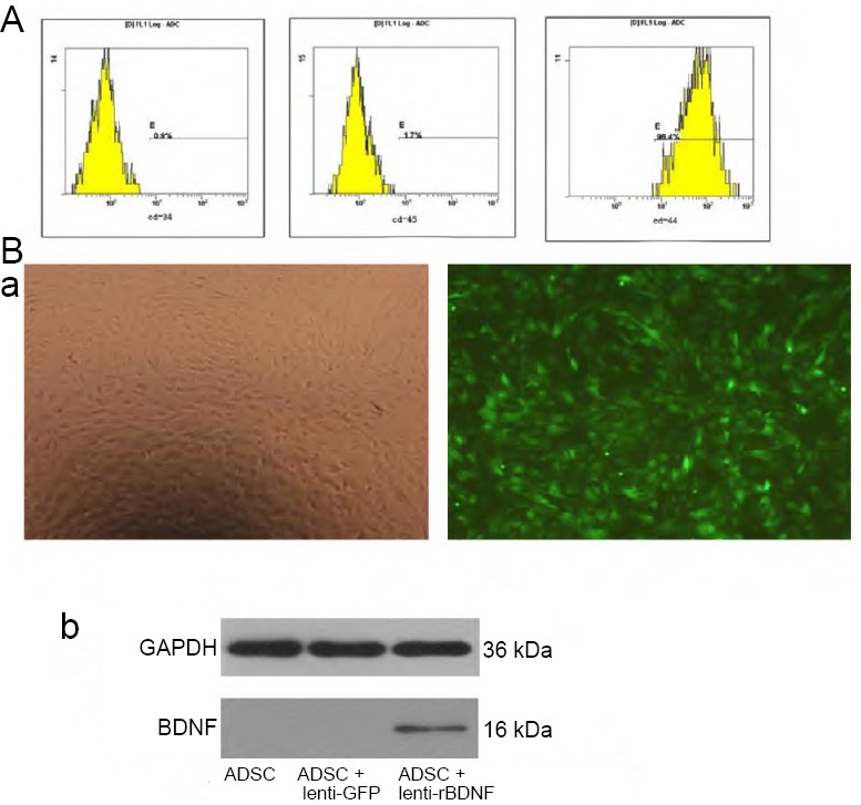
Identification and infection of ADSCs.
(A) Phenotypes of ADSCs at the third passage by flow cytometry; a: CD34 (0.9%), b: CD45 (1.7%), c: CD44 (96.4%). (B) Determination of lentiviral infection efficiency and rBDNF expression (arrows) in ADSCs. Panel (a) Infection efficiency 3 days after infection with lenti-rBDNF at an MOI of 100. GFP (green) was observed under light (left) or fluorescence (right) microscopy (original magnification, 100×). Panel (b) Representative immunoblot of rBDNF protein as measured by western blot assay. ADSCs were infected with control lentivirus (ADSC + lenti-GFP) or lenti-rBDNF (ADSC + lenti-rBDNF) at an MOI of 100. ADSCs: Adipose-derived stem cells; GFP: green fluorescent protein; MOI: multiplicity of infection; rBDNF: rat brain-derived neurotrophic factor.