
Figure 3.
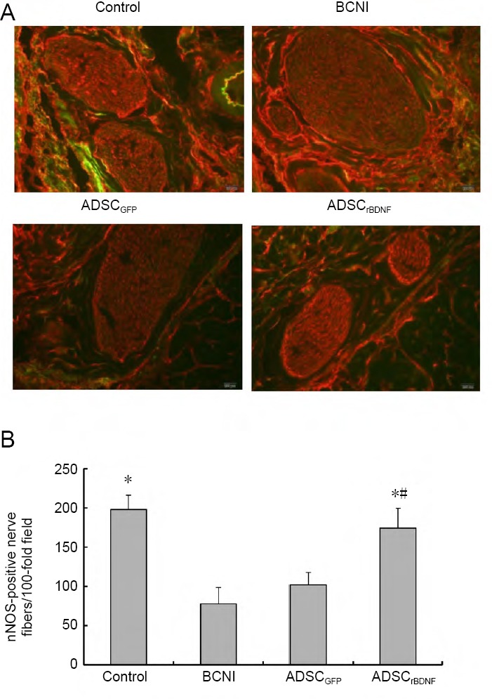
nNOS staining in a penile midshaft specimen.
(A) nNOS (red-stained fiber) in penile tissue (immunofluorescence microscope: 200×). Control group: Predominant red staining of nerve fibers; BCNI group: a paucity of red-stained fibers; ADSCrBDNF group: a significant increase in the number of red-stained nerve fibers compared with the ADSCGFP group. (B) nNOS-positive nerve fibers in penile midshaft sections. Data are expressed as the mean ± SD (n = 10; one-way analysis of variance followed by the Student-Newman-Keuls post hoc test). *P < 0.05, vs. BCNI group; #P < 0.05, vs. ADSCGFP group. ADSC: Adipose-derived stem cell; BCNI: bilateral cavernous nerve injury; GFP: green fluorescent protein; nNOS: neuronal nitric oxide synthase; rBDNF: rat brain-derived neurotrophic factor.