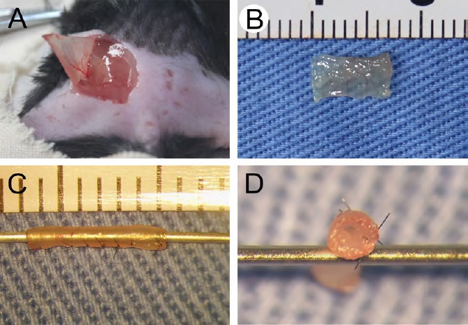
Figure 1.

Fabrication of external oblique muscle-fabricated nerve conduit.
(A) An incision was made on the dorsal region close to the tail to expose the external oblique muscle. (B) The external oblique muscle was harvested, trimmed, and cut into a 7 mm-long thin slice. (C, D) The thin skeletal muscle tissue was discontinuously sutured into a hollow conduit.
