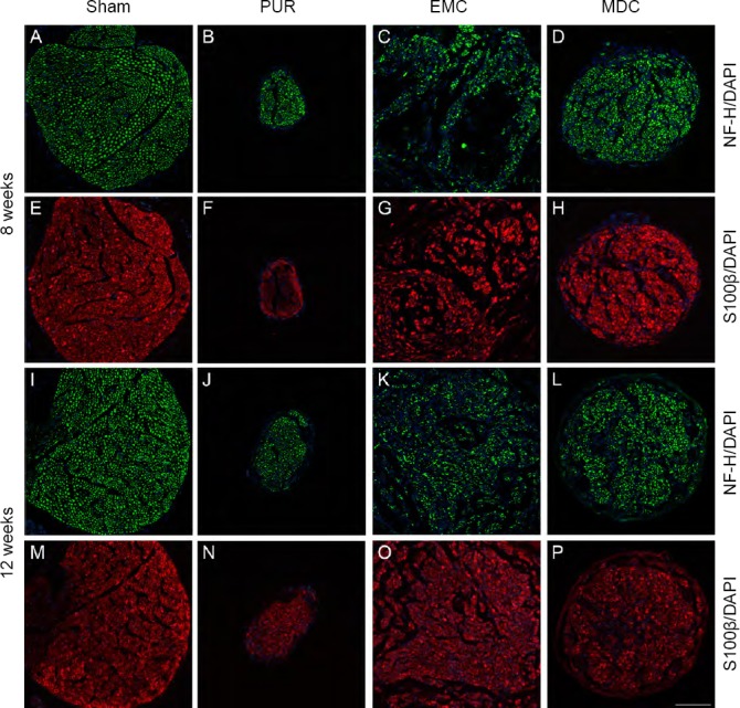
Figure 4.

Immunofluorescence staining and distribution of nerve lesions at 8 and 12 weeks post-surgery.
(A–H) Eight weeks post-surgery; (I–P) twelve weeks post-surgery. (A, E, I, M) Sham group: NF-H-expressing axons and S100β-expressing myelin sheaths were evenly distributed. (B, F, J, N) PUR group: distributions of NF-H-expressing axons and S100β-expressing myelin sheaths were uniform but with a small scope. (C, G, K, O) EMC group: distributions of NF-H-positive axons and S100β-positive myelin sheaths were not uniform at 8 weeks post-surgery (C, G), but were improved at 12 weeks post-surgery (K, O). (D, H, L, P) MDC group: NF-H-expressing axons and S100β-expressing myelin sheaths were uniformly distributed at both time points. (A–D, I–L) NF-H (green); (E–H, M–P) S100β (red); 4′,6-diamidino-2-phenylindole (DAPI; blue). Scale bar: 100 μm. DAPI: 4′,6-Diamidino-2-phenylindole; EMC: external oblique muscle-fabricated nerve conduit; MDC: muscle-derived cell; NF-H: neurofilament heavy; PUR: polyurethane; Sham: sham-operated.
