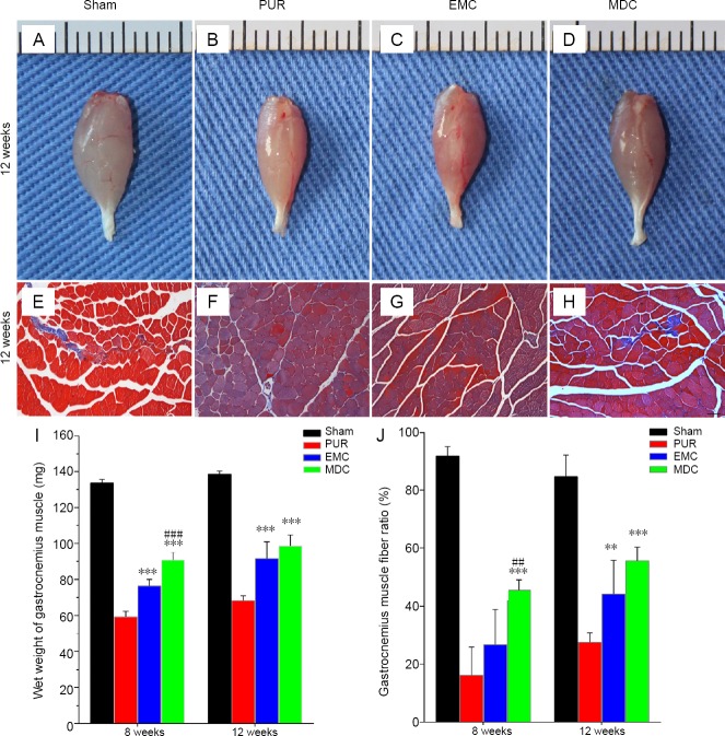
Figure 9.
General observation and Masson’s trichrome staining of gastrocnemius muscle from the repaired side at 12 weeks post-surgery, and assessment of the wet weight of gastrocnemius muscle and muscle fiber ratio at 8 and 12 weeks post-surgery.
(A–D) General observation and (E–H) cross-sections with Masson’s trichrome staining at 12 weeks after surgery. (A, E) Sham group; (B, F) PUR group; (C, G) EMC group; (D, H) MDC group. Scale bar: 100 μm. (I) Wet weight and (J) gastrocnemius muscle fiber ratio were mearsured to evaluate functional recovery of the gastrocnemius muscle. **P < 0.01, ***P < 0.001, vs. PUR group; ##P < 0.01, ###P < 0.001, vs. EMC group. Data are expressed as the mean ± SD (n = 4; one-way analysis of variance followed by Tukey’s post hoc test). EMC: External oblique muscle-fabricated nerve conduit; MDC: muscle-derived cell; PUR: polyurethane; Sham: sham-operated.

